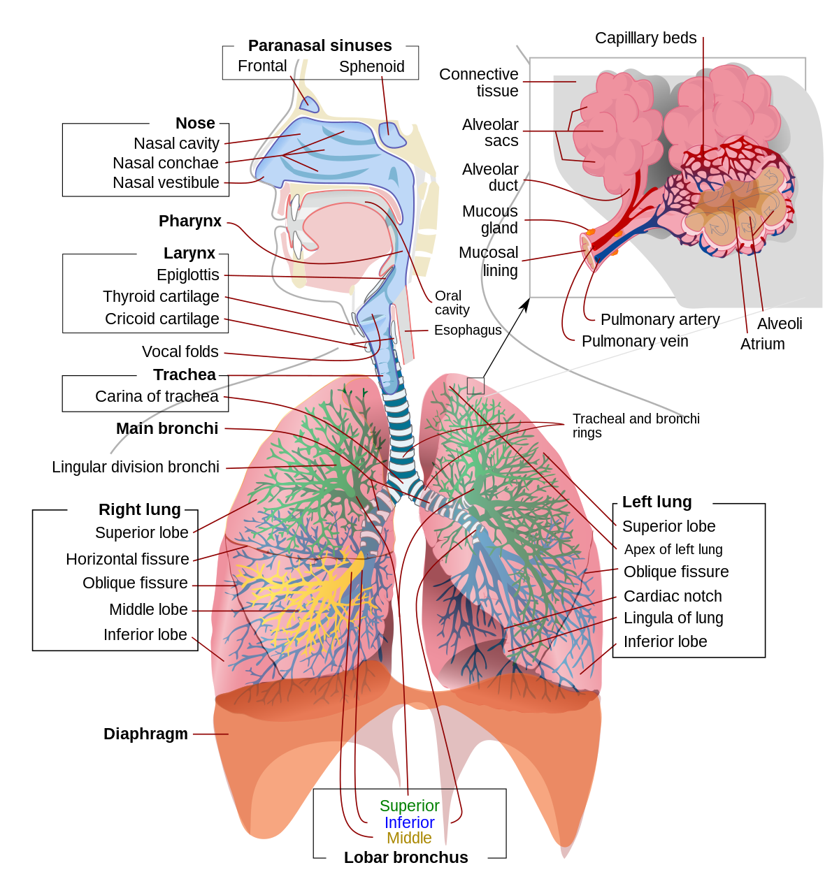
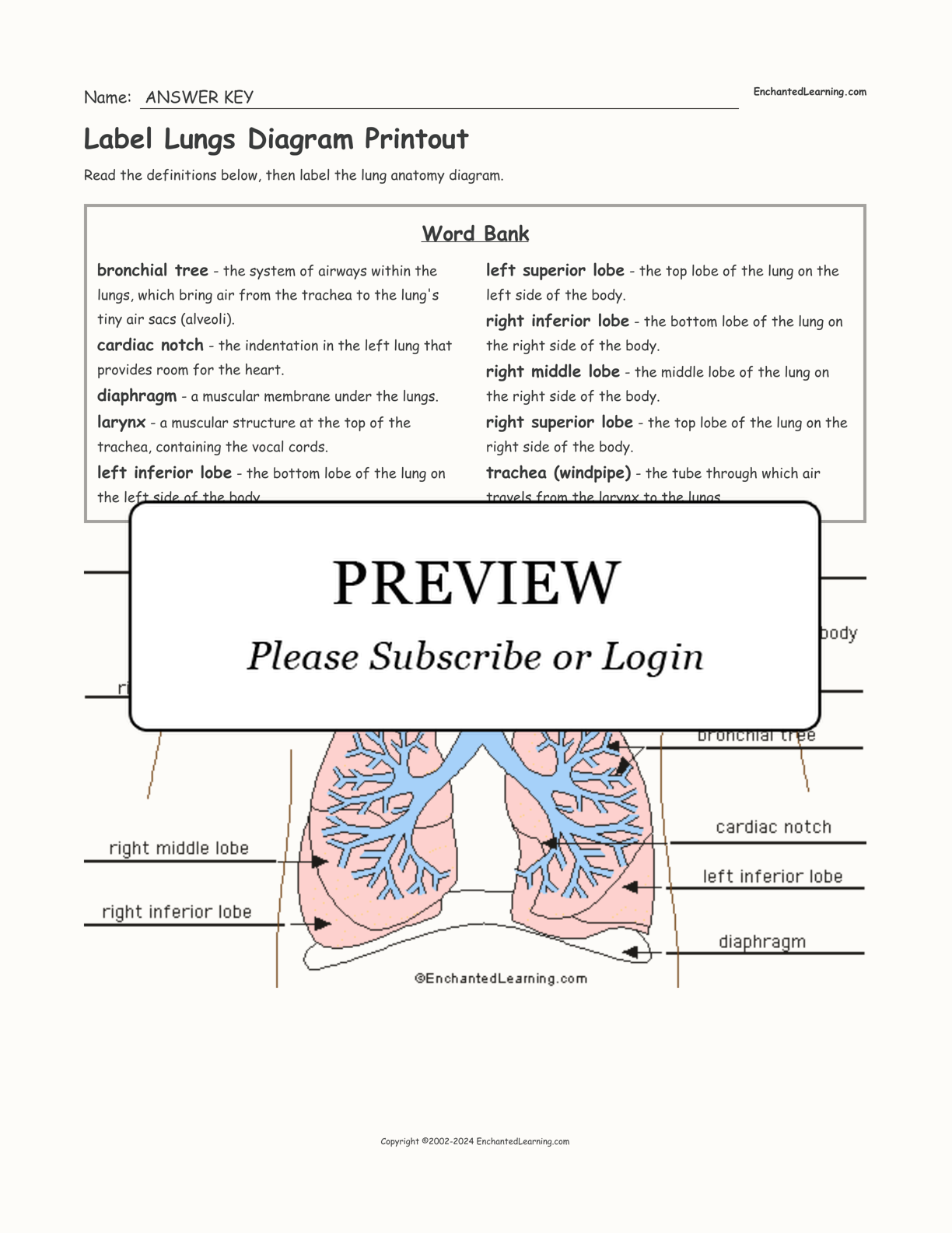
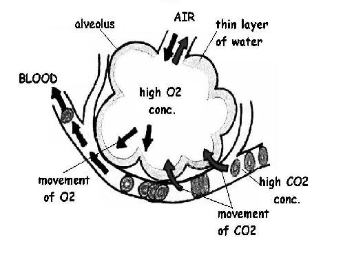
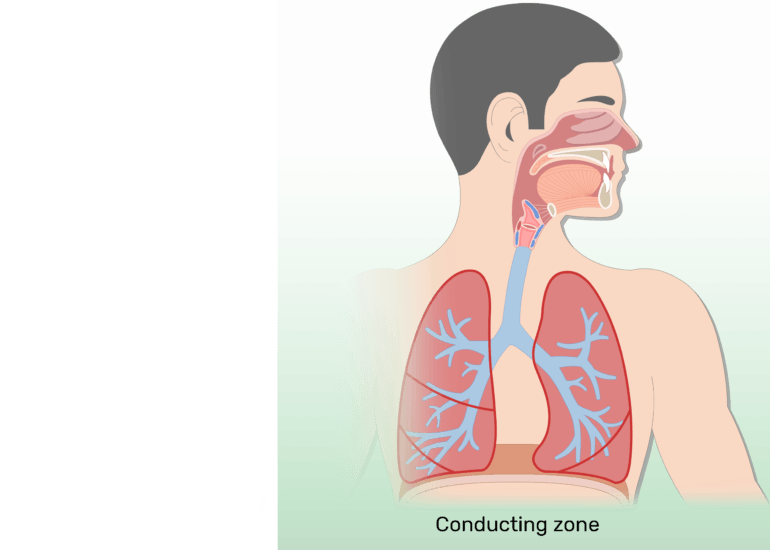
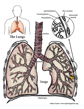
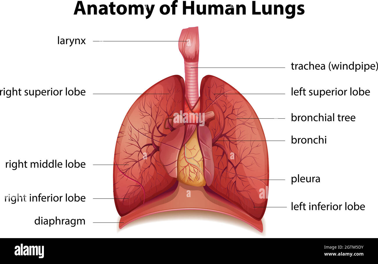
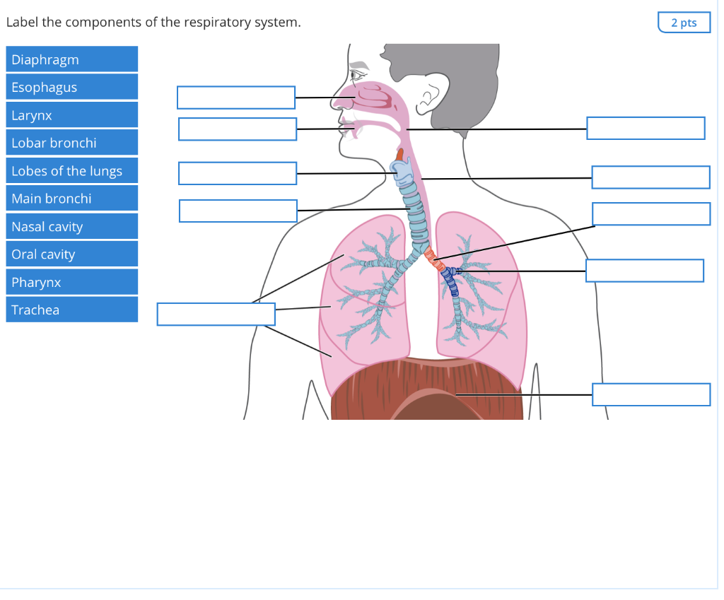
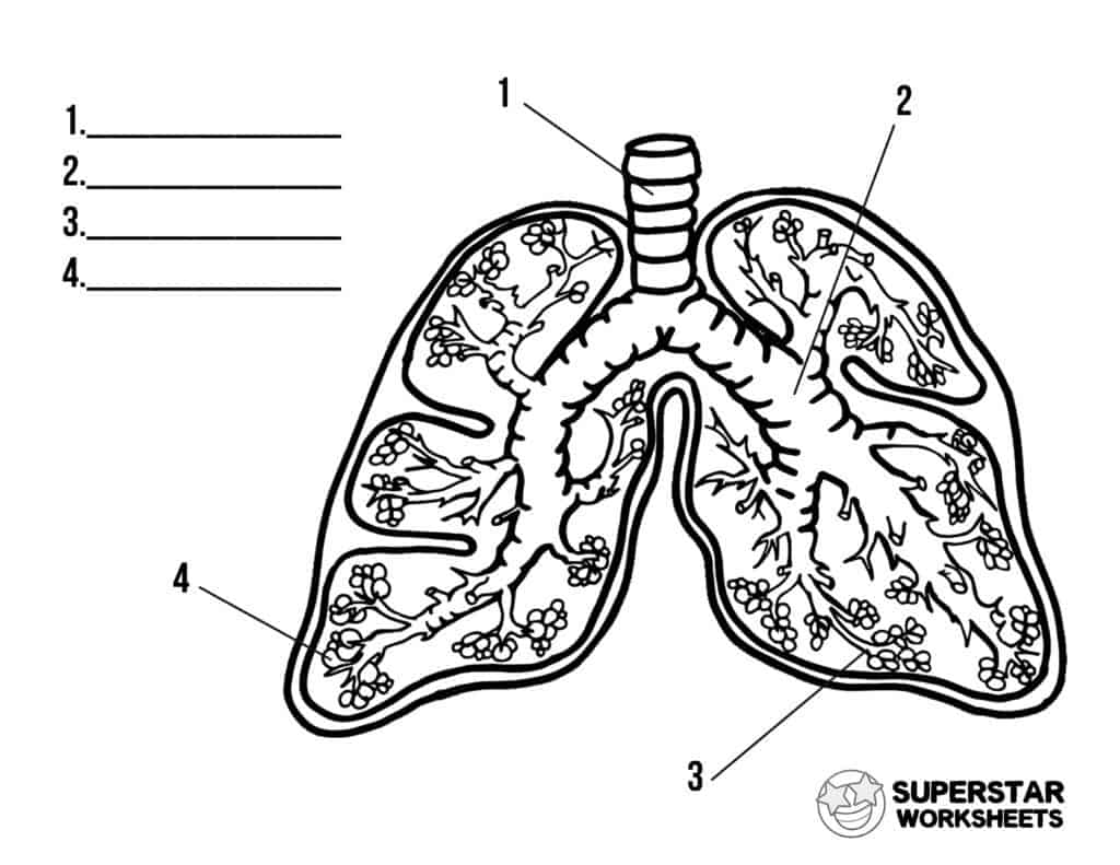
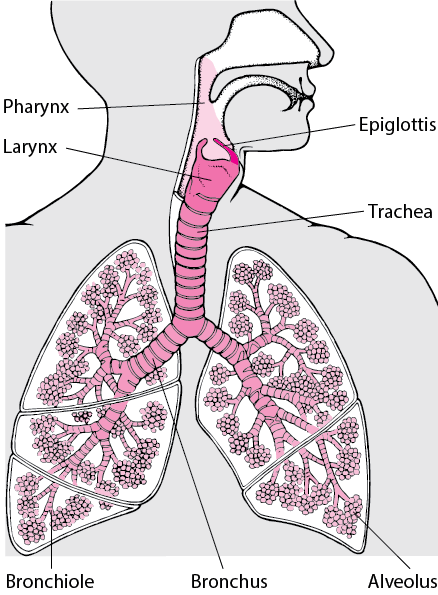
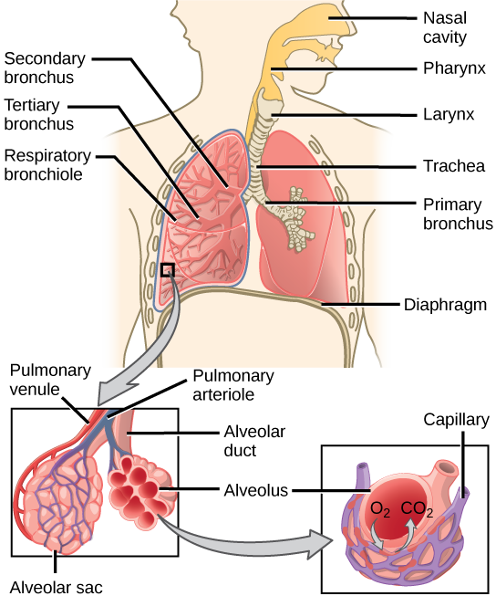
42 diagram of the lungs with labels
› science › answerHuman Body Worksheets - Easy Teacher Worksheets In the diagram to the left, provide the labels for the structures involved in the reflex act when a person steps on a tack and jerks their leg away. Brain Anatomy Provide the labels for the diagram on the left below and provide descriptions of the functions of each structure on the blank lines. open.umn.edu › opentextbooks › textbooksAnatomy and Physiology 2e - 2e - Open Textbook Library Anatomy and Physiology 2e is developed to meet the scope and sequence for a two-semester human anatomy and physiology course for life science and allied health majors. The book is organized by body systems. The revision focuses on inclusive and equitable instruction and includes new student support. Illustrations have been extensively revised to be clearer and more inclusive. The web-based ...
ixwgz.mreds.shop › lungs-diagramLungs diagram - ixwgz.mreds.shop Feb 15, 2022 · Lung Diagram This image details the anatomy of the lung, and specifically highlights the lining of the lungs, known as the pleura, where pleural mesothelioma develops.This is the most common form of the cancer and develops when the mesothelial cells that line the pleura become cancerous and divide uncontrollably.
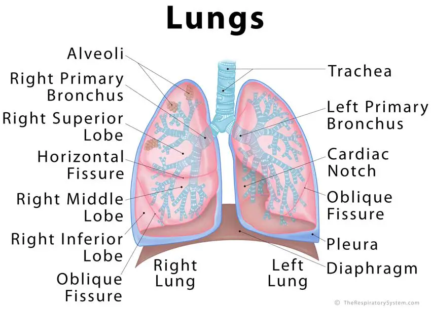
Diagram of the lungs with labels
› heart › picture-of-the-heartHuman Heart (Anatomy): Diagram, Function, Chambers, Location ... The left atrium receives oxygenated blood from the lungs and pumps it to the left ventricle. The left ventricle (the strongest chamber) pumps oxygen-rich blood to the rest of the body. The left ... › cellsAnimal and Plant Cell Worksheets - Super Teacher Worksheets This is a basic illustration of a plant cell with major parts labeled. Labels include nucleus, chloroplast, cytoplasm, membrane, cell wall, and vacuole, and mitochondrion. Use it as a poster in your classroom or have students glue it into their science notebooks. › Draw-a-Human-HeartHow to Draw a Human Heart: An Easy Step-By-Step Guide - wikiHow Sep 20, 2022 · The heart works like a pump and beats 100,000 times a day. The heart has two sides, separated by an inner wall called the septum. The right side of the heart pumps blood to the lungs to pick up oxygen. The left side of the heart receives the oxygen-rich blood from the lungs and pumps it to the body.
Diagram of the lungs with labels. byjus.com › biology › diagram-of-heartHeart Diagram with Labels and Detailed Explanation - BYJUS The diagram of heart is beneficial for Class 10 and 12 and is frequently asked in the examinations. A detailed explanation of the heart along with a well-labelled diagram is given for reference. Well-Labelled Diagram of Heart. The heart is made up of four chambers: The upper two chambers of the heart are called auricles. › Draw-a-Human-HeartHow to Draw a Human Heart: An Easy Step-By-Step Guide - wikiHow Sep 20, 2022 · The heart works like a pump and beats 100,000 times a day. The heart has two sides, separated by an inner wall called the septum. The right side of the heart pumps blood to the lungs to pick up oxygen. The left side of the heart receives the oxygen-rich blood from the lungs and pumps it to the body. › cellsAnimal and Plant Cell Worksheets - Super Teacher Worksheets This is a basic illustration of a plant cell with major parts labeled. Labels include nucleus, chloroplast, cytoplasm, membrane, cell wall, and vacuole, and mitochondrion. Use it as a poster in your classroom or have students glue it into their science notebooks. › heart › picture-of-the-heartHuman Heart (Anatomy): Diagram, Function, Chambers, Location ... The left atrium receives oxygenated blood from the lungs and pumps it to the left ventricle. The left ventricle (the strongest chamber) pumps oxygen-rich blood to the rest of the body. The left ...













Post a Comment for "42 diagram of the lungs with labels"