40 brain mri with labels
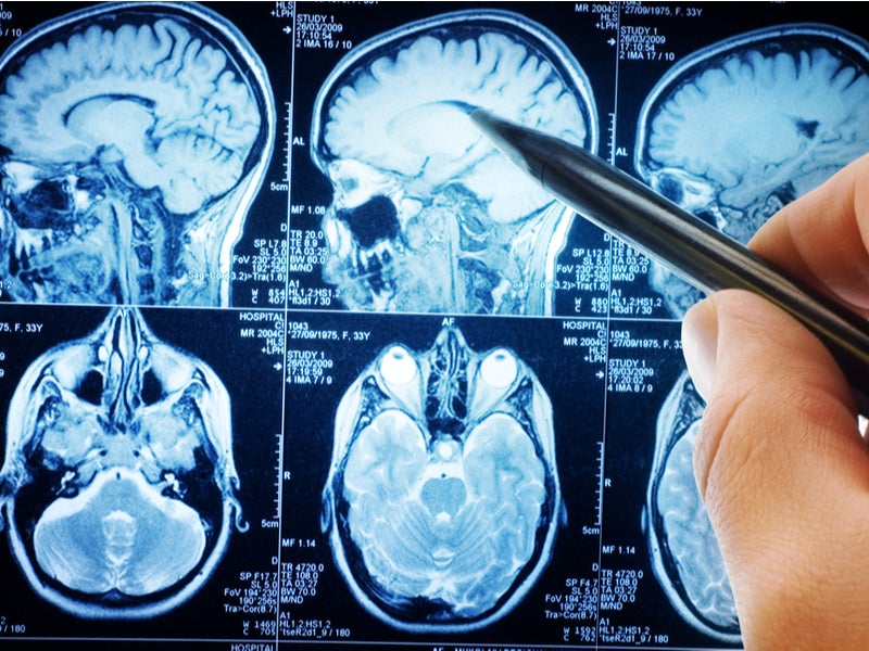
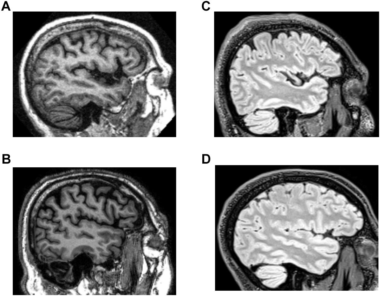
› 2013 › 07CPT Code for MRI Brain, Breast, Lumbar Spine and Shoulder Find below the latest Radiology CPT codes for for MRI of Brain, Breast, Lumbar Spine and Shoulder: CPT Codes for MRI Lumbar spine In human Lumbar spine is represented by the 5 vertebrae in between the ribcage and the pelvis forming the largest segment of the vertebral column. Depending on the condition that one is treated on these parts of the ... IMAIOS An MRI was performed on a healthy subject, with several acquisitions with different weightings: spin-echo T1, T2 and FLAIR, T2 gradient-echo, diffusion, and T1 after gadolinium injection. We obtained 24 axial slices of the normal brain.
Automatic labelling of brain tissues in MR images through spatial ... To reduce the cost of manual operations and make it possible to label with a large number of 3D MR images, the automatic labelling methods for brain anatomical structures have been developed to find a general model to guide the segmentation, such as the segmentation of subcortical brain structures using fuzzy templates , probabilistic atlases ...

Brain mri with labels
Labeling Brain Structures - John Muschelli In Processing Within-Visit MRI we show how to register a T1-weighted image to the Eve template. The Eve template has two full brain segmentations and one white matter segmentations, each done by hand. I will refer to these as "atlases" because they tell you "where" you are in the brain with the corresponding labels. 1 Labels in template space Magnetic Resonance Imaging (MRI) of the Spine and Brain Magnetic resonance imaging (MRI) is a diagnostic procedure that uses a combination of a large magnet, radiofrequencies, and a computer to produce detailed images of organs and structures within the body. Unlike X-rays or computed tomography (CT scans), MRI does not use ionizing radiation. Some MRI machines look like narrow tunnels, while others ... Classification of brain tumours in MR images using deep ... - Nature A brain tumour is the growth of abnormal cells in the brain. Brain tumours are classified based on their speed of growth and the likeness of them growing back after treatment. They are mainly...
Brain mri with labels. Arterial Spin Labeling Perfusion of the Brain: Emerging Clinical ... Introduction. Arterial spin labeling (ASL) is a magnetic resonance (MR) imaging technique that enables the measurement of brain perfusion noninvasively at the tissue level.Benefiting from the contrast of inflowing magnetically labeled blood, ASL obviates an exogenous contrast agent. Although the principle of ASL was introduced in early 1990s (1-3) and is feasible on low-field-strength MR ... Brain Tumor MRI Classification | VGG16 | Kaggle Brain Tumor MRI Classification | VGG16. Python · Keras Pretrained models, Brain MRI Images for Brain Tumor Detection. Labels · Mobashra/Brain-MRI-Tumor-Classification · GitHub Contribute to Mobashra/Brain-MRI-Tumor-Classification development by creating an account on GitHub. 101 labeled brain images and a consistent human cortical labeling ... We introduce the Mindboggle-101 dataset, the largest and most complete set of free, publicly accessible, manually labeled human brain images. To manually label the macroscopic anatomy in magnetic resonance images of 101 healthy participants, we created a new cortical labeling protocol that relies on robust anatomical landmarks and minimal manual edits after initialization with automated labels.
MRI anatomy | free MRI axial brain anatomy - Mrimaster.com WRIST CORONAL. KNEE CORONAL. KNEE SAGITTAL. ARTERIES UPPER LEG. ARTERIES LOWER LEG. This MRI brain cross sectional anatomy tool is absolutely free to use. Use the mouse scroll wheel to move the images up and down alternatively use the tiny arrows (>>) on both side of the image to move the images. Researchers automate brain MRI image labeling, more than ... - ScienceDaily Researchers have automated brain MRI image labeling, needed to teach machine learning image recognition models, by deriving important labels from radiology reports and accurately assigning them to ... › AANLIB › casesHarvard University Show labels Show list All modalities to: MR-T1 MR-T2 FDG T1/FDG T2/FDG Brain: Atlas of human anatomy with MRI - e-Anatomy - IMAIOS The module on the anatomy of the brain based on MRI with axial slices was redesigned, having received multiple requests from users for coronal and sagittal slices. The elaboration of this new module, its labeling of more than 524 structures on 379 MRI images in three different views and on 26 anatomical diagrams, took more than 6 months.
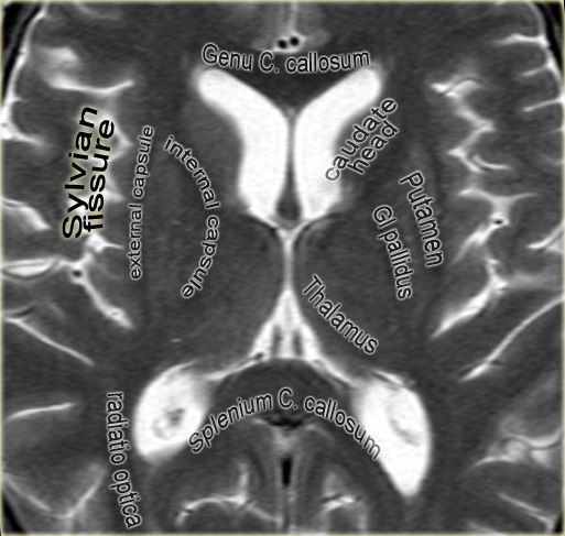
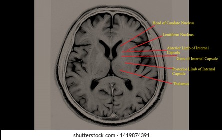
Arterial spin labeling MRI: clinical applications in the brain Arterial spin labeling (ASL) perfusion magnetic resonance imaging (MRI) sequences are increasingly being used to provide MR-based CBF quantification without the need for contrast adm … Visualization of cerebral blood flow (CBF) has become an important part of neuroimaging for a wide range of diseases. Brain MRI: How to read MRI brain scan | Kenhub The insular and limbic lobes are the ones of particular interest in the brain MRI. The insular lobe lies just lateral to the extreme capsule of basal ganglia. It is a small portion of the cerebral cortex found deep to the meeting point of the frontal, temporal and parietal lobes. The limbic lobe lies deep to the parietal and frontal lobes. It is a functional unit often referred to as the limbic system. Atlas of BRAIN MRI - W-Radiology Brain magnetic resonance imaging (MRI) is a common medical imaging method that allows clinicians to examine the brain's anatomy (1). It uses a magnetic field and radio waves to produce detailed images of the brain and the brainstem to detect various conditions (2). Automated MRI image labelling processes 100,000 brain exams in under 30 ... Researchers from the School of Biomedical Engineering & Imaging Sciences at King's College London have automated brain MRI image labeling, needed to teach machine learning image recognition models,...
Head and spine anatomy - Radiology Cafe
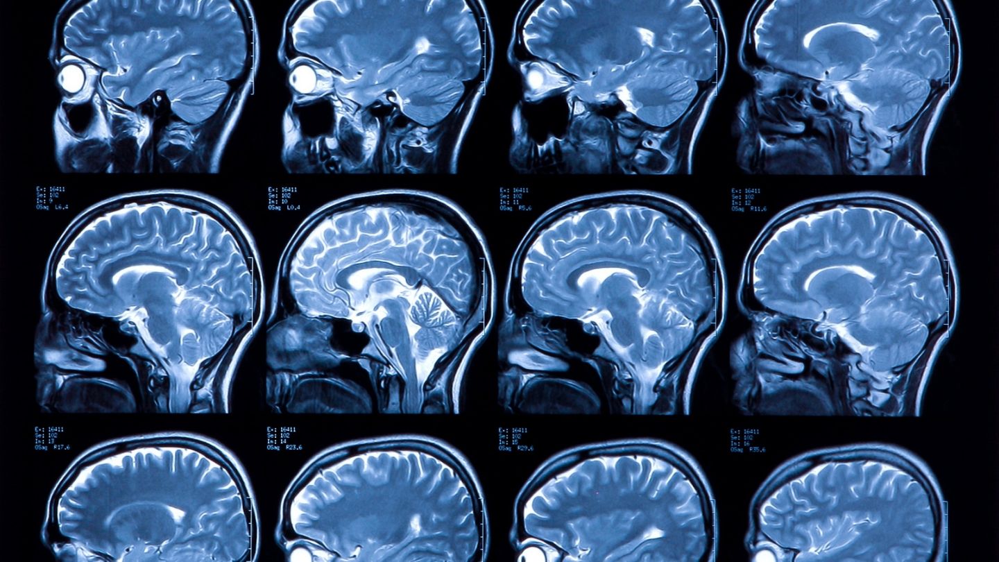
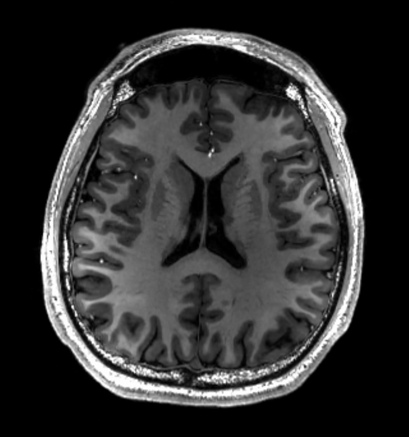
Normal brain (MRI) | Radiology Case | Radiopaedia.org MRI Axial T2 Normal appearance of a young person's brain on a 1.5T scanner other than borderline low-lying tonsils. Note, however, that McRae's line (basion to the opisthion) needs to be measured A) in the midline and B) from the tip of the cortical bone - and not the fat-rich bone marrow.
MRI scans prove useful for understanding depression
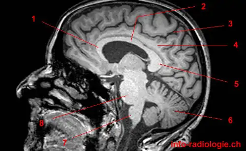
Brain lobes - annotated MRI | Radiology Case | Radiopaedia.org Brain by Dr Roya Faghani; IMPORTANTS by Dr Ahmed Faiz Al-Musawi; atlantic fellow session by Ophir Keret; Brain Anatomy by Dr Renor Gonçalves de Castro Neto brain anatomy by Diana Paduraru; Year 2 Investigation Session - Neuroradiology (Cases) by Dr Sally Ayesa Neuro by Ravikiran; anatomy by Tayson Nguyen; Labeled Anatomy by Abdullah Abohimed

MRI head axial T2 - labeling questions | Radiology Case ...
Enhancing the REMBRANDT MRI collection with expert segmentation labels ... The board-certified radiologist performed labeling of the MRI scans of the all modalities in the dataset that included MRIs from different modalities, including T1-weighted, T2-weighted,...
Potentially life-saving study could cut labelling times for ...
medicalxpress.com › news › 2022-08-poor-heart-healthPoor heart health predicts premature brain ageing Aug 22, 2022 · More information: Life course, genetic, and neuropathological associations with brain age in the British 1946 birth cohort: a population-based study, The Lancet Healthy Longevity (2022).DOI: 10. ...

MRI anatomy | free MRI axial brain anatomy
Automatic anatomical brain MRI segmentation combining label propagation ... In this study, we examined the performance and processes of label propagation with decision fusion in MR images of the human brain, starting from detailed expert segmentations of 30 brains and using a registration method based on free-form deformation (Rueckert et al., 1999). The aim of this work was to evaluate the accuracy of the method by ...
Atlas of BRAIN MRI - W-Radiology
PDF Learning-based 3T brain MRI segmentation with guidance from 7T MRI labeling Key words: segmentation, brain MRI, 7T MRI labeling, high magnetic field 1. INTRODUCTION Magnetic resonance imaging (MRI) is a powerful tool for in vivo diagnosis of brain disorders. Accurate measurement of brain structures in MRI is important for studying both brain development associated with growth and brain alterations associated with ...
The Radiology Assistant : Anatomy
101 Labeled Brain Images and a Consistent Human Cortical Labeling Protocol In this article we introduce this dataset of manually edited brain image labels applied to the T1-weighted MR images of publicly available multi-modal data acquired from healthy individuals. We also introduce a benchmark for the evaluation of automated registration/segmentation/labeling methods by comparing the manual labels according to this "Desikan-Killiany-Tourville" (DKT) protocol with automatically generated labels.

Approach to MRI brain | LearningNeurology.com
Automatic labeling and classification of brain CT images In this way, the ROIs are automatically labeled with pathology types which can be served as class labels, and a training data set of a large number of training instances is generated automatically....
Basal Ganglia Annotated Structures Brain Mri Stock ...
› view › transdermal-patchesTransdermal Patches That Must Be Removed Before MRI Aug 26, 2016 · Although MRI’s capabilities are well-recognized, its inherent dangers can’t be overlooked. One report from the Joint Commission showed burns are the most common injuries in the MRI suite. 2 The most common objects to undergo significant heating are wires and leads, pulse oximeter sensors and cables, and safety pins and metal clamps.
Magnetic resonance imaging of the brain - Wikipedia
A neuroradiologist's guide to arterial spin labeling MRI in clinical ... Abstract. Arterial spin labeling (ASL) is a non-invasive MRI technique to measure cerebral blood flow (CBF). This review provides a practical guide and overview of the clinical applications of ASL of the brain, as well its potential pitfalls. The technical and physiological background is also addressed.

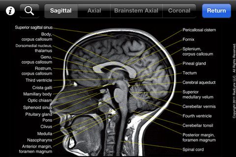
Brain: Atlas of human anatomy with MRI - e-Anatomy
› en › e-AnatomyShoulder: MRI, radiographical, and illustrated anatomical ... Sep 13, 2021 · MRI of the shoulder : muscles of the rotator cuff labeled on a sagittal MR slice. An MRI of the shoulder of a healthy subject was performed in the 3 planes of space (coronal, axial, sagittal) commonly used in osteoarticular imaging, with two weightings to explore the musculoskeletal pathology of the shoulder: spin-echo T1 and proton-density ...

brain anatomy | MRI coronal brain anatomy | free MRI cross ...
Arterial spin labeling MRI: Clinical applications in the brain Arterial spin labeling (ASL) is a completely noninvasive magnetic resonance imaging (MRI) method that uses magnetically labeled blood water as a flow tracer, providing CBF images of the brain. Moreover, if certain conditions are met it can potentially also provide an absolute, quantifiable CBF measurement on a voxel-by-voxel basis 2.
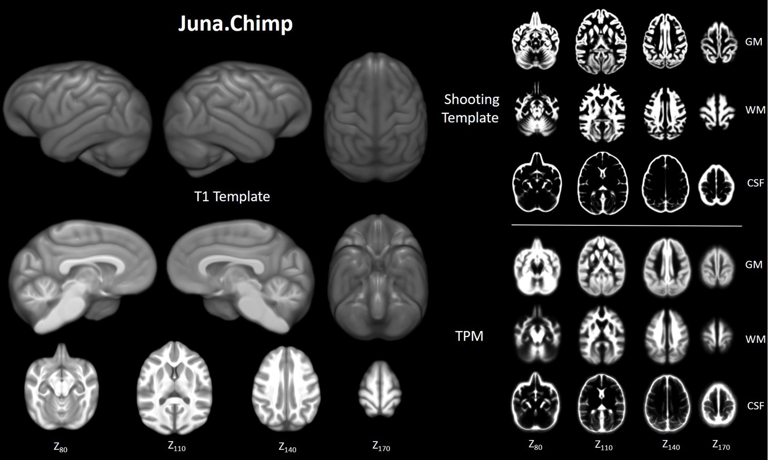
Chimpanzee brain morphometry utilizing standardized MRI ...
› en › e-AnatomyNormal chest MDCT with anatomic labels | e-Anatomy - IMAIOS Mar 10, 2022 · IMAIOS and selected third parties, use cookies or similar technologies, in particular for audience measurement. Cookies allow us to analyze and store information such as the characteristics of your device as well as certain personal data (e.g., IP addresses, navigation, usage or geolocation data, unique identifiers).

Neural Structure Quiz
NITRC: Manually Labeled MRI Brain Scan Database: Tool/Resource Info Manually Labeled MRI Brain Scan Database. Visit Website. Image 1 of 3. Click for more. This is a continuously growing and improving database of high-quality neuroanatomically labeled MRI brain scans, created not by an algorithm, but by neuroanatomical experts. All results are checked and corrected. Regions of interest include the usual sub ...

How much does a brain MRI cost? | From $225
Frontiers | Brain Tumor MR Image Classification Using Convolutional ... Brain tumor image classification is an important part of medical image processing. It assists doctors to make accurate diagnosis and treatment plans. Magnetic resonance (MR) imaging is one of the main imaging tools to study brain tissue. In this article, we propose a brain tumor MR image classification method using convolutional dictionary learning with local constraint (CDLLC). Our method ...
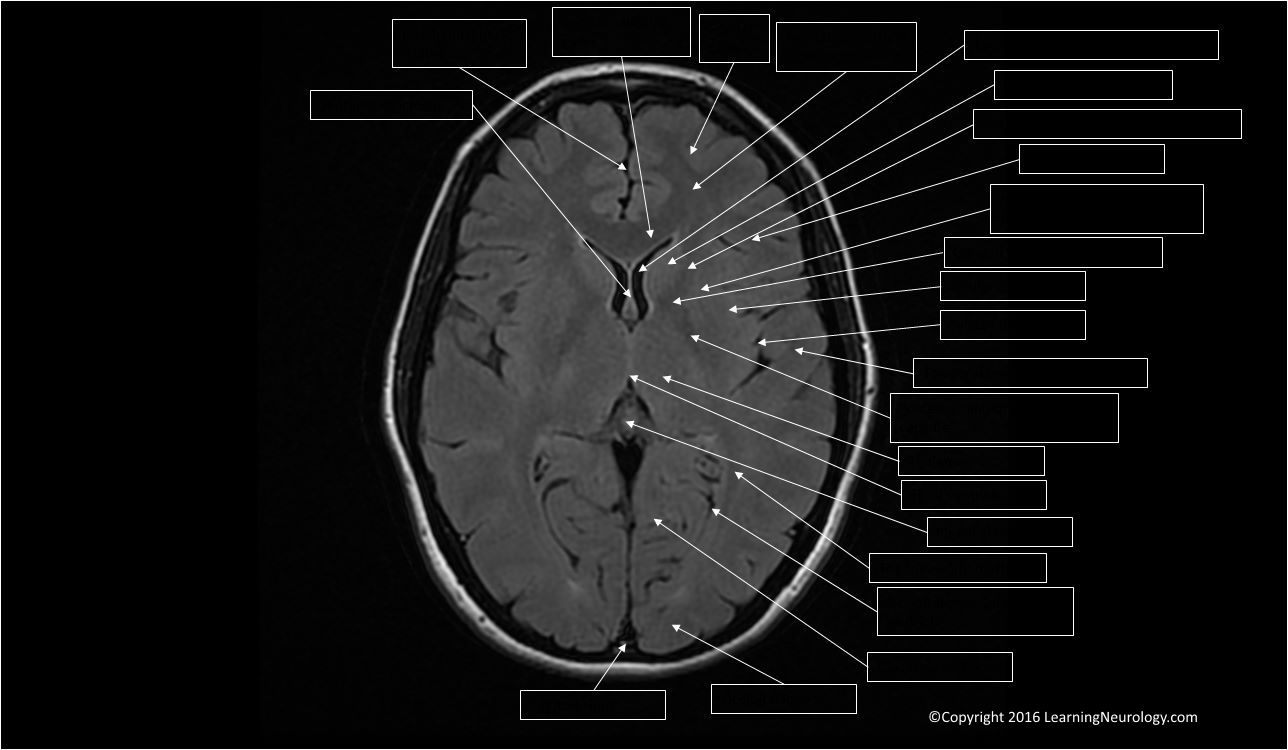
Approach to MRI brain | LearningNeurology.com
› articles › s41586/022/04554-yBrain charts for the human lifespan | Nature Apr 06, 2022 · To extend the scope of brain charts beyond the four cerebrum tissue volumes, we generalized the same GAMLSS modelling approach to estimate normative trajectories for additional MRI phenotypes ...
Orientation and Voxel-Order Terminology: RAS, LAS, LPI, RPI ...
Brain MRI segmentation | Kaggle This dataset contains brain MR images together with manual FLAIR abnormality segmentation masks. The images were obtained from The Cancer Imaging Archive (TCIA). They correspond to 110 patients included in The Cancer Genome Atlas (TCGA) lower-grade glioma collection with at least fluid-attenuated inversion recovery (FLAIR) sequence and genomic cluster data available.

Whole-Brain Arterial Spin Labeling Perfusion MRI in Patients ...
Classification of brain tumours in MR images using deep ... - Nature A brain tumour is the growth of abnormal cells in the brain. Brain tumours are classified based on their speed of growth and the likeness of them growing back after treatment. They are mainly...

Brain Tumor Detection and Localization - Analytics Vidhya
Magnetic Resonance Imaging (MRI) of the Spine and Brain Magnetic resonance imaging (MRI) is a diagnostic procedure that uses a combination of a large magnet, radiofrequencies, and a computer to produce detailed images of organs and structures within the body. Unlike X-rays or computed tomography (CT scans), MRI does not use ionizing radiation. Some MRI machines look like narrow tunnels, while others ...
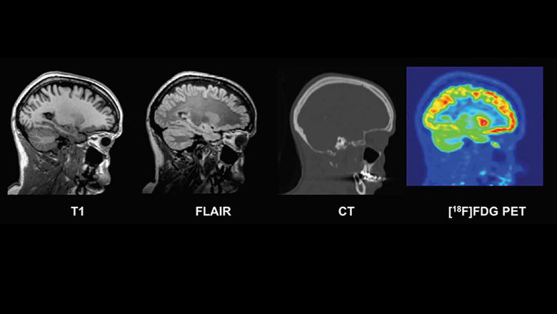
New database of healthy adult human brain PET, MRI and CT ...
Labeling Brain Structures - John Muschelli In Processing Within-Visit MRI we show how to register a T1-weighted image to the Eve template. The Eve template has two full brain segmentations and one white matter segmentations, each done by hand. I will refer to these as "atlases" because they tell you "where" you are in the brain with the corresponding labels. 1 Labels in template space

Delaware Neuroscience - Brain Bee Detail, Page 2

Labelled MRI of Normal Brain - Stock Image - C017/4418 ...

Intelligent Scanning Using Deep Learning for MRI — The ...

The role of computed tomography (CT) and magnetic resonance ...

Tips and traps in brain MRI: Applications to vascular ...

MRI anatomy | free MRI axial brain anatomy

MRI Segmentation of the Human Brain: Challenges, Methods, and ...

Magnetic Resonance Imaging (MRI): Brain - Connecticut Children's

MRI anatomy | free MRI axial brain anatomy

Normal anatomy of the brain on CT and MRI with a few normal ...
Frontiers | Systematic Differences Between Perceptually ...

Gulf View Medical Centre - Did you know MRI's are commonly ...

Cross sectional Anatomy of Brain on... - World Of Radiology ...
MRI identifies markers of atypical brain deve | EurekAlert!

CerebrA: Accurate registration and manual label correction of ...

MRI anatomy | free MRI axial brain anatomy
Judith Shirley (judithshirley96) – Profile | Pinterest

7.0 T MRI Axial Images. (a) An axial view image obtained by ...
Magnetic resonance images of the brain (MRI brain) sagittal ...

Anatomy of the brain (MRI) | Mri brain, Anatomy, Mri
Post a Comment for "40 brain mri with labels"