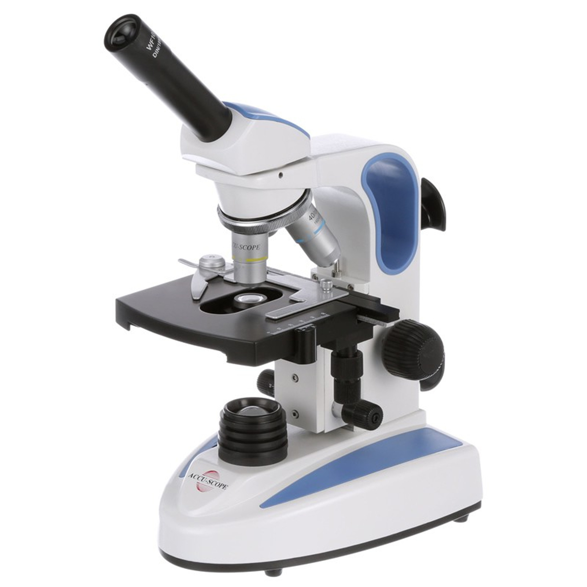
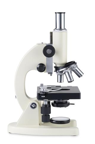
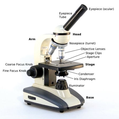
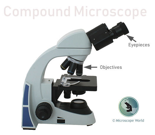
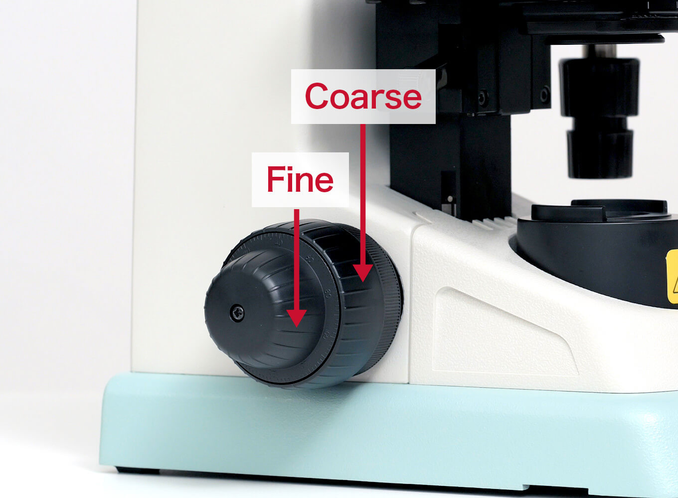
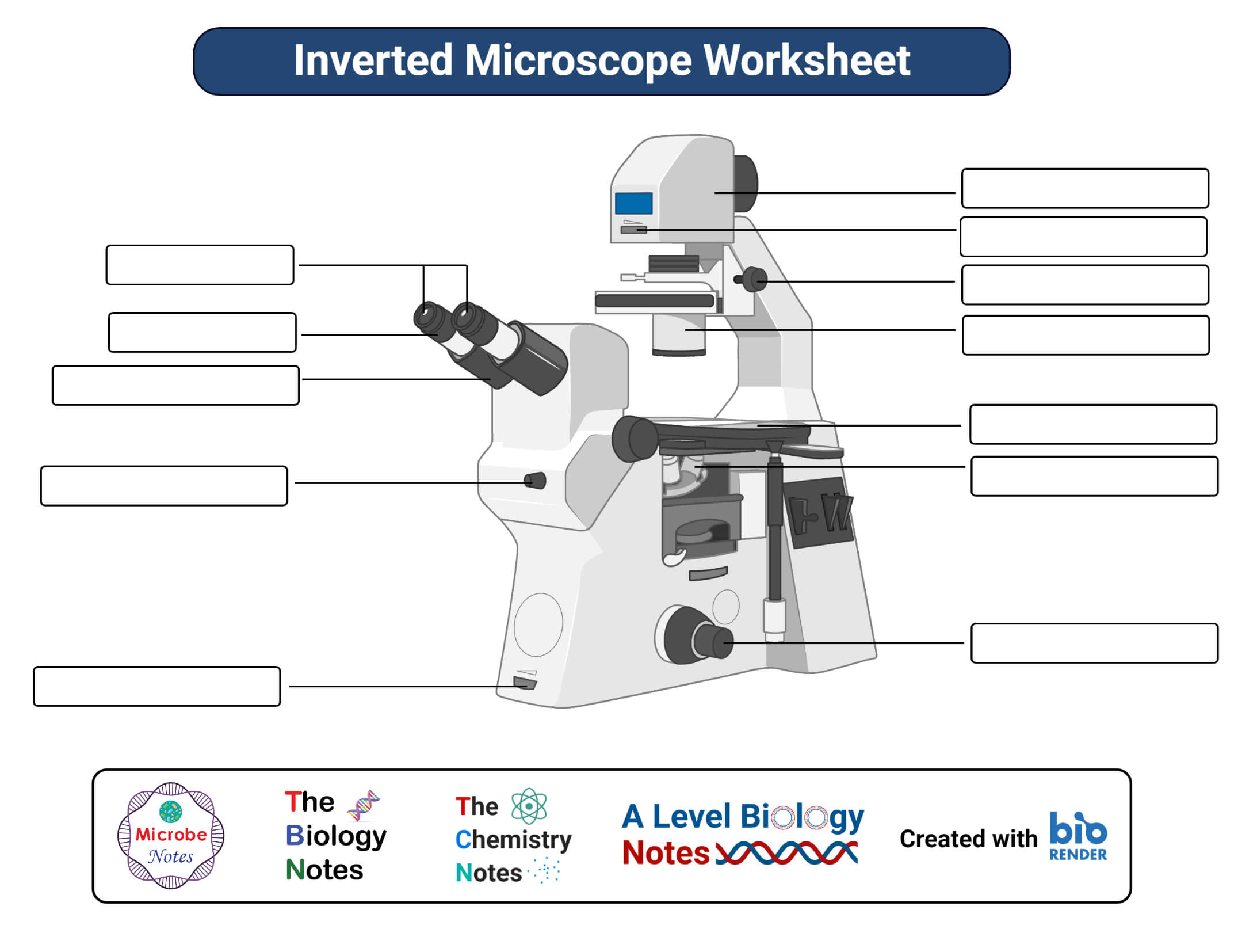
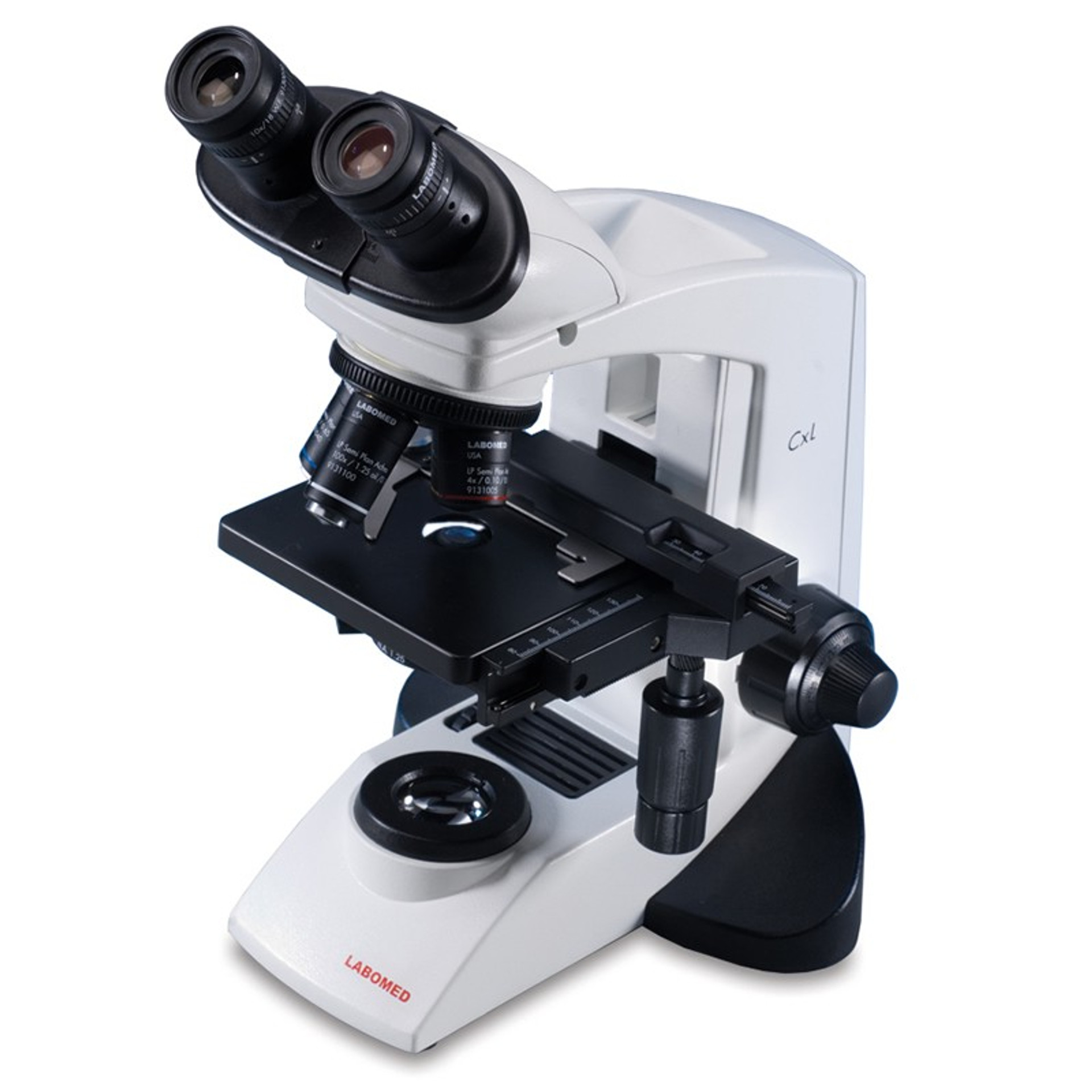
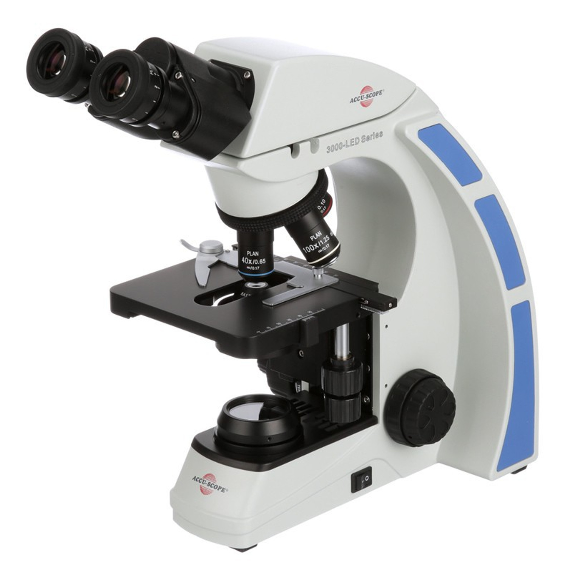
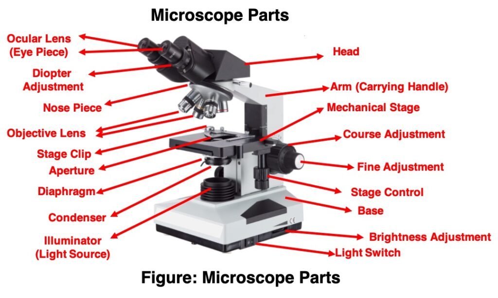
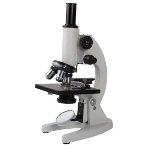
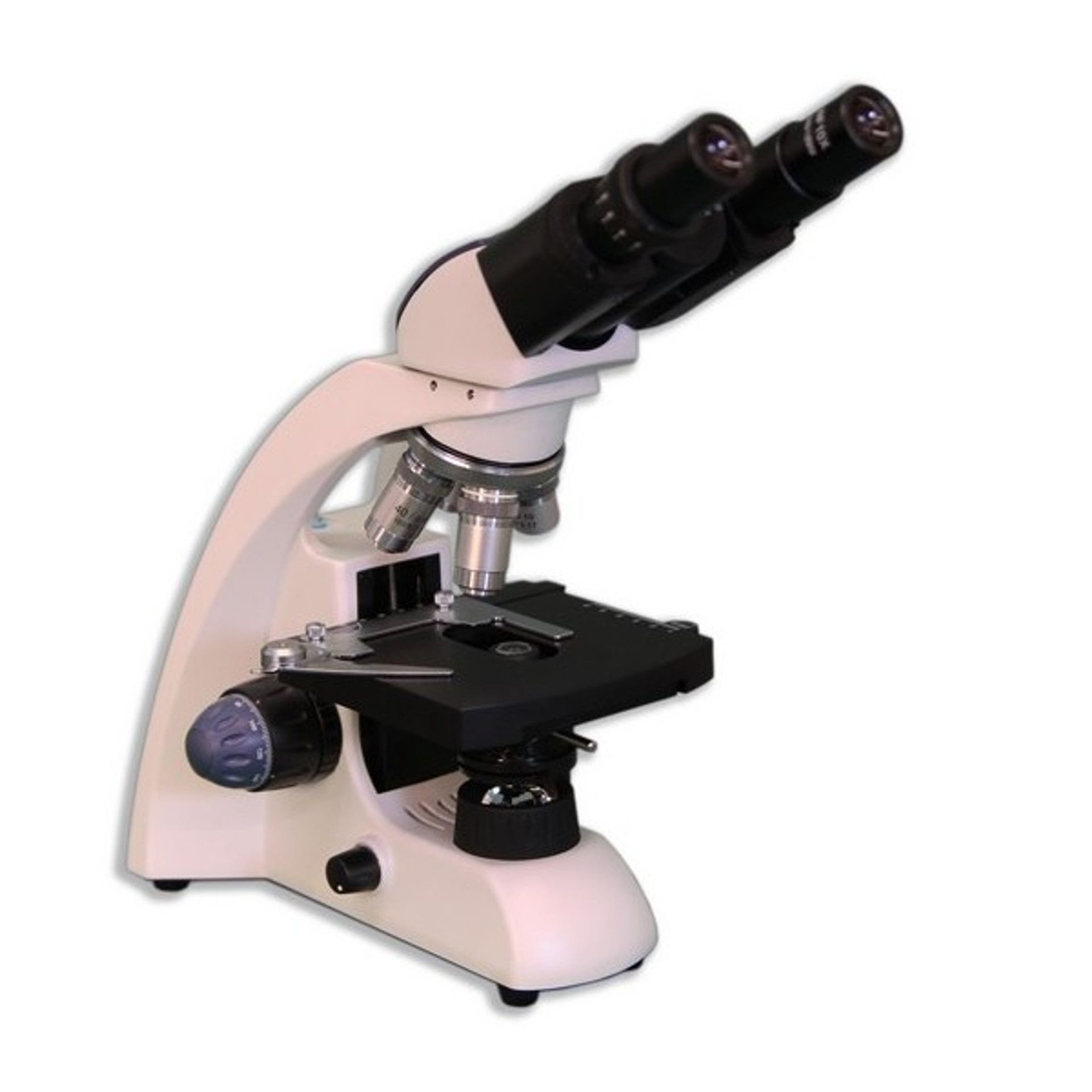
39 labels of a microscope and functions
Robert Hooke - Biography, Facts and Pictures Robert Hooke’s own illustration of his compound microscope, with labels added by this website. Hooke used his microscope to observe the smallest, previously hidden details of the natural world. His book Micrographia revealed and described his discoveries. Some people disputed his diagrams because they refused to believe what they showed. The world Hooke had discovered … Electron microscope - Wikipedia An electron microscope is a microscope that uses a beam of accelerated electrons as a source of illumination. As the wavelength of an electron can be up to 100,000 times shorter than that of visible light photons, electron microscopes have a higher resolving power than light microscopes and can reveal the structure of smaller objects.. Electron microscopes use shaped magnetic …
Histology - Yale University Bone is a tissue in which the extracellular matrix has been hardened to accommodate a supporting function. The fundamental components of bone, like all connective tissues, are cells and matrix. There are three key cells of bone tissue. They each have unique functions and are derived from two different cell lines.

Labels of a microscope and functions
Parts of Stereo Microscope (Dissecting microscope) – labeled … Compared to a compound microscope where the objectives attached to the nosepiece can be seen and identified individually (based on color bands and their respective labels), the objectives of a dissecting microscope are located in a cylindrical cone and, therefore, are not directly seen. For the stereo microscope that comes with multiple objective lens sets (fixed power style), the … Label the microscope — Science Learning Hub 08.06.2018 · All microscopes share features in common. In this interactive, you can label the different parts of a microscope. Use this with the Microscope parts activity to help students identify and label the main parts of a microscope and then describe their functions.. Drag and drop the text labels onto the microscope diagram. If you want to redo an answer, click on the … A FINE GEORGE II MAHOGANY CASED CUFF PATTERN MONOCULAR MICROSCOPE A FINE GEORGE II MAHOGANY CASED CUFF PATTERN MONOCULAR MICROSCOPEJOHN CUFF, LONDON, MID 18th CENTURYThe body tube with stepped moulded shuttered eyepiece over ogee waist and objective tube incorporating marks for six positions on an exponential scale numbered 1 to 6, supported via a tapered collar set in a ring attached to a vertical slide moving against the fixed limb upright marked with six ...
Labels of a microscope and functions. (PDF) Introduction to Microscopy - ResearchGate 08.11.2017 · This new microscope doesn't require any special labels and could help increase access to low-cost plant science diagnostic tool. The hands on training of the microscopic instrument generally ... The Contrast Transfer Function - Image Formation | Coursera 14.12.2019 · Just like in the previous slide where we had two different wave functions that had the same wavelength. But they were phase shifted with respect to each other slightly. And because of that, some of them added significantly to the sum and others did not add significantly to the sum. Whether they add or not, this is dependent on the phase shifts that are created by … LAS X Industry Microscope software for Industry | Products Activate all relevant functions (e.g. for illumination settings, camera, measurements) with a few clicks ; Automatically store images on a regular basis with functionalities like autosave; Customizable user access. The software can handle multiple users who have different levels of microscope skills and diverse tasks to accomplish. Profiles according to user’s skills. The LAS … Introduction to three-dimensional image processing Introduction to three-dimensional image processing¶. Images are represented as numpy arrays. A single-channel, or grayscale, image is a 2D matrix of pixel intensities of shape (row, column).We can construct a 3D volume as a series of 2D planes, giving 3D images the shape (plane, row, column).Multichannel data adds a channel dimension in the final position containing color …
The Brain - Science Quiz - GeoGuessr The Brain - Science Quiz: The term “gray matter” is an unflattering term for such an important organ, but that’s what the brain looks like—a gray blob. That blob is actually a sophisticated neural command center for the entire body, controlling all functions and enabling our thoughts, emotions, fears, and dreams. This science quiz game will help you learn the 7 parts of the brain. A FINE GEORGE II MAHOGANY CASED CUFF PATTERN MONOCULAR MICROSCOPE A FINE GEORGE II MAHOGANY CASED CUFF PATTERN MONOCULAR MICROSCOPEJOHN CUFF, LONDON, MID 18th CENTURYThe body tube with stepped moulded shuttered eyepiece over ogee waist and objective tube incorporating marks for six positions on an exponential scale numbered 1 to 6, supported via a tapered collar set in a ring attached to a vertical slide moving against the fixed limb upright marked with six ... Label the microscope — Science Learning Hub 08.06.2018 · All microscopes share features in common. In this interactive, you can label the different parts of a microscope. Use this with the Microscope parts activity to help students identify and label the main parts of a microscope and then describe their functions.. Drag and drop the text labels onto the microscope diagram. If you want to redo an answer, click on the … Parts of Stereo Microscope (Dissecting microscope) – labeled … Compared to a compound microscope where the objectives attached to the nosepiece can be seen and identified individually (based on color bands and their respective labels), the objectives of a dissecting microscope are located in a cylindrical cone and, therefore, are not directly seen. For the stereo microscope that comes with multiple objective lens sets (fixed power style), the …










Post a Comment for "39 labels of a microscope and functions"